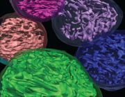
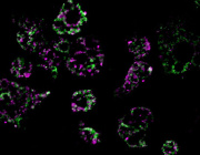
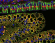
Novel movies showing lipid droplets and mitochondria fine dynamics live and at high resolution


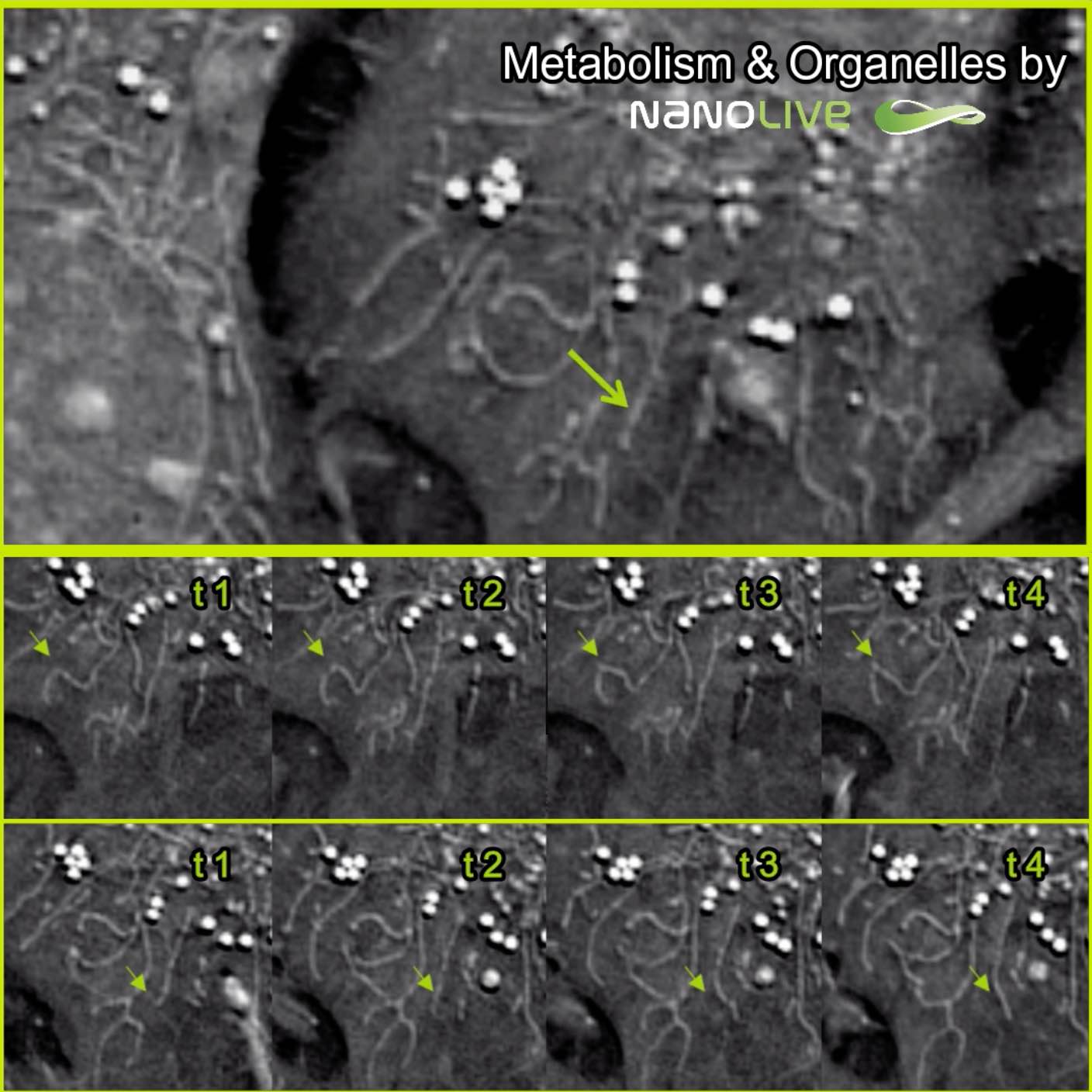
Thanks to the 3D Cell Explorer’s live imaging capabilities, highlights of lipid droplets and mitochondria fine dynamics that were previously out of reach are clearly visible now.
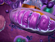
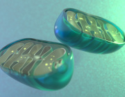
On the top panel you can observe a time-lapse video of of pre-adipocytes imaged with the 3D Cell Explorer for 1 hour at a frequency of one image per five seconds (movie speed: 15fps). On the middle panel are displayed four time point images of a mitochondrial fission happening in the cell. On the bottom panel a fusion process is displayed.
During the Targeting Mitochondria 2019 Congress, Holotomographic Phase Imaging or short Nanolive Imaging will be presented at the Targeting Mitochondria with a talk and booth.
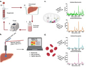
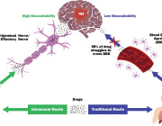
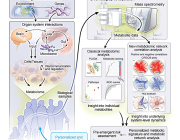
Non-invasive live cell imaging overcomes phototoxicity problem while imaging cellular and mitochondrial processes
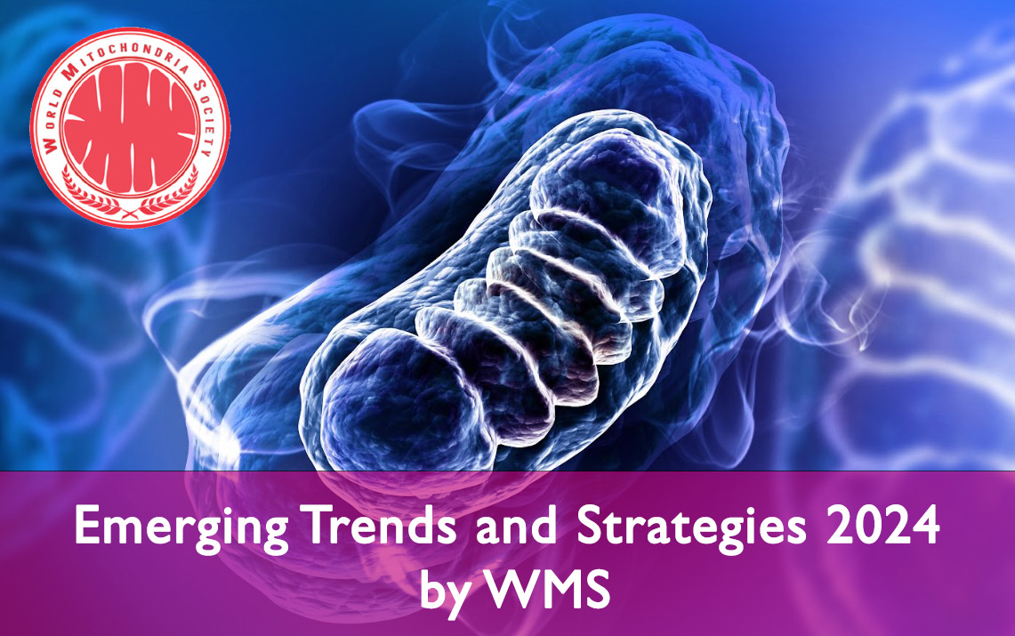
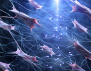
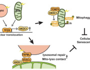
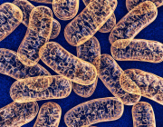
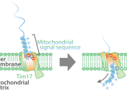
Mitochondria are key organelles for various essential cellular processes.
A better understanding of the dynamics and interactions with other organelles could benefit research as well as future therapeutic approaches. The Targeting Mitochondria 2019 will host a broad spectrum of speakers advancing our common scientific field. The introduction of a new technology for label free gentle live cell imaging of Mitochondria might add an important new instrument to this advancement. Holotomographic Phase Imaging or short Nanolive Imaging will be presented at the Targeting Mitochondria with a talk and booth.
A major problem with current imaging techniques is phototoxicity that leads to the observation of perturbed dynamics. Consequently, the mitigation of phototoxicity leads to poor time resolution of time lapse approaches. This is particularly true for small organelles like mitochondria or lipid droplets that are extremely sensitive to photo-induced oxidation. Last but not least, the use of chemical or genetically-encoded fluorescent markers perturbs the targeted biological processes.
However, the Nanolive Imaging allows visualisation of Mitochondria without any staining or labelling. Further, it requireds ~100 times less energy (~0.2 nW/µm2) than light sheet microscopes (~1nW/µm2). With a resolution below 200 nm, it enables high resolution and high-frequency imaging even with sensitive material, giving access to organelle dynamics that were previously out of reach.
Please follow this link to Watch the video.
10th Anniversary of Targeting Mitochondria Congress
October 28-29, 2019 - Berlin, Germany
www.mitochondria-site.com